
Untitled. Alveolar Epithelial Cells Are Critical in Protection of the Respiratory Tract by Secretion of Factors Able To Modulate the Activity of Pulmonary Macrophages and Directly Control Bacterial Growth. The respiratory epithelium is a physical and functional barrier actively involved in the clearance of environmental agents.
The alveolar compartment is lined with membranous pneumocytes, known as type I alveolar epithelial cells (AEC I), and granular pneumocytes, type II alveolar epithelial cells (AEC II). AEC II are responsible for epithelial reparation upon injury and ion transport and are very active immunologically, contributing to lung defense by secreting antimicrobial factors. AEC II also secrete a broad variety of factors, such as cytokines and chemokines, involved in activation and differentiation of immune cells and are able to present antigen to specific T cells. Another cell type important in lung defense is the pulmonary macrophage (PuM). Considering the architecture of the alveoli, a good communication between the external and the internal compartments is crucial to mount effective responses.
Fig 1 Fig 2 Additionally, we tested medium obtained from spleen cells and LPS. Untitled. Areolar Tissue - Structure and Functions of Human Tissue Types. Note: This page is part of the section about the structure and function of different Tissue Types, which is related to the section about Histology and Cells (incl. structure of animal cells, cell division, mitosis, meiosis).
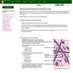
This "Tissue Types" section is included to complete description of the knowledge of "Histology - The Cell" required by some courses in First-Level Anatomy and Physiology. To read about other tissue types see the list of on the left. Areolar Tissue is a common form of loose connective tissue. 1.0 Where in the body is Areolar tissue ? Areolar tissue is found in many locations around the body. 2.0 The Structure of Areolar tissue. What are some types of respiratory system tissue? Anatomy and Physiology: The Relationships of the Respiratory System. Place your hand over your chest, take a deep breath, and then let it out.
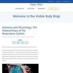
Of course you already know that your lungs fill with air when you breathe, but did you know that your respiratory system does more than simply move oxygen into and out of your lungs? The structures of the respiratory system interact with structures of the skeletal, circulatory, and muscular systems to help you smell, speak, and move oxygen into your bloodstream and waste out of it.
We're going to take a look at the relationships between these systems and how they work to keep you breathing 24/7/365. The Lungs of the Respiratory System The lungs are asymmetrical, conical in shape, and have a spongy texture. Did you know the surface area of one lung is 750 sq. feet? Air flows from the trachea into the bronchi, and from there into the bronchioles of the lungs.
Standard Grade Bitesize Physical Education - Structure and function : Revision, Page 5. Diaphragm - Anatomy Pictures and Information. [Continued from above] . . .

The lungs are enclosed in the thoracic cavity by the rib cage on the front, back, and sides with the diaphragm forming the floor of the cavity. When we inhale, the diaphragm contracts and is drawn inferiorly into the abdominal cavity until it is flat. At the same time, the external intercostal muscles between the ribs elevate the anterior rib cage like the handle of a bucket.
The thoracic cavity becomes deeper and larger, drawing in air from the atmosphere. During exhalation, the rib cage drops to its resting position while the diaphragm relaxes and elevates to its dome-shaped position in the thorax. Structurally, the diaphragm consists of two parts: the peripheral muscle and central tendon. The peripheral muscle can be further divided by its origins into the sternal, costal, and lumbar regions. Prepared by Tim Taylor, Anatomy and Physiology Instructor. Interactive Anatomy Guide. The cells of the human body require a constant stream of oxygen to stay alive.
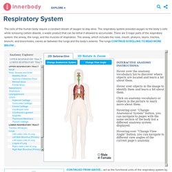
The respiratory system provides oxygen to the body’s cells while removing carbon dioxide, a waste product that can be lethal if allowed to accumulate. Untitled. Untitled.