
Microfracture Procedure Rehabilitation. Michael M.
Reinold, PT, DPT, SCS, ATC, CSCS; Kevin E. Wilk, PT, DPT This protocol for rehabilitation after knee microfracture surgery is designed to provide the rehabilitation professional with a general guideline for patient care with the AlterG Anti-Gravity Treadmill. As such, it should be stressed that this is only a protocol and should not be a substitute for professional clinical decision-making regarding a patient’s progression.
And it should be further noted that progression should be individualized based upon each patient’s specific needs, surgical variables, pain level, the specific surgeon’s guidelines, physical examination, progress, and presence of any complications. Download Protocol for printing. Week 0 – Week 4 Goals: Protect healing tissue from load and shear forces Decrease pain and effusion Restoration of full passive knee extension Gradually restore knee flexion Regain quadriceps control Reduce edema & pain. Rehabilitation following microfracture surgery. Much of the success of microfracture surgery for articular cartilage lesions in the knee depends on what happens after the surgery is over.

Progressive, controlled loading of the repaired joint is the key to safe and effective rehabilitation. By Jon Fravel, ATC, and Michael Shaffer PT, ATC, OCS Injuries to the articular cartilage of the knee are serious, with potentially debilitating consequences if not managed appropriately. Articular cartilage injuries can occur from either an acute traumatic event or from chronic degeneration.1 Acute traumatic injuries producing cartilage lesions are most often seen in a younger athletic population, while chronic degenerative lesions most often occur in older individuals. Articular cartilage injuries are diagnosed both by clinical exam and imaging. Cartilage lesions occur across a spectrum of injury. Stephanie E. Siegrist, MD. M0455763. Vastus_Medialis_Oblique_Weakness_NMES. Electrical Muscle Stimulation. From Running tips for everyone from beginners to racing marathons and ultramarathons Using EMS on the VMO.
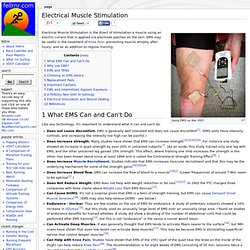
Electrical Muscle Stimulation is the direct of stimulation a muscle using an electric current that is applied via electrode patches on the skin. EMS may be useful in the treatment of Knee Pain, preventing muscle atrophy after injury, and as an addition to regular training. Like any technology, it's important to understand what it can and can't do. Does not cause discomfort. The main reasons to use EMS are around injury treatment and rehabilitation. EMS is similar to TENS (Transcutaneous Electrical Nerve Stimulation), and many devices, such as the one recommended below, a capable of both EMS and TENS.
EMS devices vary wildly in price, from less than $50 to over $800. Max current: ~100mA Frequency range: 1-150Hz Pulse width: 50-400us Wave Form: Square Wave. Different devices had two to eight electrodes (one to four channels), and some devices had a TENS mode for pain reduction. EMS FOR THE VASTUS MEDIALIS OBLIQUUS. Electrical muscle stimulation for the vastus medialis obliquus muscle (VMO) can work wonders in most knee problems.
It can also be used as to help prevent knee problems, and to protect the hips What you need: 1. The muscle stimulator. AustJPhysiotherv32i4McConnell. Chondromalacia Patella Grade 3. . . Quote: Well, it's hard to say.
There is research that shows being active is the best way to maintain function and joint space in individuals with signs of osteoarthritis (essentially where you heading with a Grade III chrondromalacia). However, excessive volume or intensity of loading can cause further degeneration. I don't have a research article that suggests either way for someone in your situation. Essentially, it's a decision that you'll have to make. If I were you, I would probably not do heavy weightlifting in high degrees of knee flexion as I mentioned before. Personally, I wouldn't pursue a marathon. You could supplement part of your training on the elliptical, as you said, as it may avoid very sharp increases in quad activity, which create sudden bursts of patellofemoral joint contact and pressure. The medical world is still trying to find a good way to restore cartilage. Patellofemoral Pain Syndrome.
From Physiopedia Definition/Description Patellofemoral Pain Syndrome (PFPS) is an umbrella term used for pain arising from the patellofemotal joint itself, or adjacent soft tissues.
Historicaly it has bee referred to as anterior knee pain but this is misleading as the pain can be felt in all aspects of the knee, (including the popliteal fossa) Be aware that PFPS is sometimes confused with chondromalacia patellae (which is a cartilage problem) and patellar tendonitis. Patients will (in general) complain about similar symptoms but the cause is different and so is the treatment. Clinically Relevant Anatomy The knee (art.
The Best Exercise for Patellofemoral Pain Syndrome. Before I get to the exercise, I got a few videos for you.
Approximately 60% of athletes have patellofemoral pain syndrome (PFPS) sometime in their life, and a long line of research has shown that PFPS is primarily caused by muscle imbalances in the vastus medialis oblique (VMO) and vastus lateralis (VL) muscles. Activation, endurance and strengthening of these muscles is key to PFPS prevention and rehabilitation, but the best exercises for these muscles have not been conclusively determined. In an effort to help clarify contradictory findings in the exercise science literature on this topic, researchers in the United Kingdom conducted a study designed to test the effect of two closed kinetic chain exercises and one open kinetic chain exercise on VMO and VL muscle activity in healthy individuals.
Highlights of the Study The study’s participants were 11 men and 11 women between the ages of 18 and 40 who were not experiencing any symptoms of PFPS at the time of the study. Exercises They Tested. 6946.pdf. Patellofemoral-Chondromalacia.