
Lee Banting
Senior Lecturer in Medicinal Chemsitry
CH391L/S12/SpinachRNA. From OpenWetWare The ‘Spinach’ aptamer is a short RNA sequence recently developed in the lab of Samie R.

Jaffrey (lab website) at Cornell University. The aptamer is capable of mimicking the fluorescence properties of EGFP (Enhanced Green Fluorescent Protein) when in complex with its ligand. Spinach derives its name from the bright green color of fluorescence and its potential utility. Fluorescence is actually a property of the aptamers ligand, which is strongly and specifically induced by specific binding of the aptamer [1]. While the benefits of GFP for studying proteins are obvious to anyone working in the various fields in biology, no analogous tool has ever been developed or discovered for RNA. Spinach Development of Spinach During structural maturation, the Green Fluorescent Protein (GFP) chromophore is formed by the covalent cyclization of three amino acids (Ser65-Tyr66-Gly67) to form a molecule known as 4-hydroxybenzlidine imidazolinone (HBI). For fish, fear smells like sugar.
When one fish gets injured, the rest of the school takes off in fear, tipped off by a mysterious substance known as "Schreckstoff" (meaning "scary stuff" in German).

Now, researchers reporting online on February 23 in the Cell Press journal Current Biology have figured out what that scary stuff is really made of. Within that chemical brew is a special type of sugar found in abundance in fish skin. When a fish is wounded, fragments of the sugar known as chondroitin sulfate alarm other fish nearby. "Our results provide a solution to a 70-year-old puzzle: the nature of this alarm signal," says Suresh Jesuthasan of A*Star's Neuroscience Research Partnership and the Duke/National University of Singapore Graduate Medical School in Singapore.
Chondroitin sulfate is a major component of fish skin, he said, and its breakdown -- likely triggered by enzymes released upon injury -- seems to be the key. The researchers suggest that those neurons may be specialized to detect the sugary alarm cue. Ron Eglash on African fractals.
Current Computer Modeling Cannot Explain Why Two Highly Similar Sequences Fold into Different Structures - Biochemistry. Laboratory of Physical Chemistry, Swiss Federal Institute of Technology ETH, 8093 Zürich, Switzerland Biochemistry, 2011, 50 (50), pp 10965–10973 DOI: 10.1021/bi2015663 Publication Date (Web): November 14, 2011 Copyright © 2011 American Chemical Society Funding Statement This work was supported by the National Center of Competence in Research (NCCR) in Structural Biology, by Grant 200020-121913 from the Swiss National Science Foundation, and by Grant 228076 from the European Research Council.

Section: Abstract The remarkable recent creation of two proteins that fold into two completely different and stable structures, exhibit different functions, yet differ by only a few amino acids poses a conundrum to those hoping to understand how sequence encodes structure. Citing Articles View all 7 citing articles. The accumulation of un-repairable DNA damage in laminopathy progeria fibroblasts is caused by ROS generation and is prevented by treatment with N-acetyl cysteine.
+ Author Affiliations ↵*To whom correspondence should be addressed.

Tel: +44 1913341270; Fax: +44 1913341201; Email: c.j.hutchison@durham.ac.uk Received May 17, 2011. Accepted July 20, 2011. Abstract Fibroblasts from patients with the severe laminopathy diseases, restrictive dermopathy (RD) and Hutchinson Gilford progeria syndrome (HGPS), are characterized by poor growth in culture, the presence of abnormally shaped nuclei and the accumulation of DNA double-strand breaks (DSB). Laminopathies are a group of inherited degenerative disorders that are caused by mutations in the gene LMNA.
Lamin A is first synthesized as a precursor molecule-termed prelamin A, which then undergoes a sequence of post-translational processing steps. It has recently been shown that cells and tissues from mouse models of HGPS, as well as fibroblasts from HGPS, RD and MAD patients accumulate DNA double-strand breaks (DSB) and are sensitized to ionizing radiation (15–18). Figure 1. Figure 2. Parallels Plesk Panel 9.5.3 for Linux. Remember The Milk: Online to do list and task management. Ferrofluid. Ferrofluid on glass, with a magnet underneath.
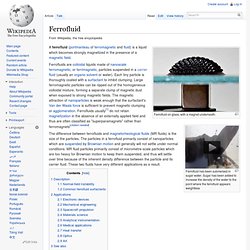
Ferrofluid has been submersed in sugar water. Sugar has been added to increase the density of the water to the point where the ferrofluid appears weightless A ferrofluid (portmanteau of ferromagnetic and fluid) is a liquid which becomes strongly magnetized in the presence of a magnetic field. Ferrofluids are colloidal liquids made of nanoscale ferromagnetic, or ferrimagnetic, particles suspended in a carrier fluid (usually an organic solvent or water). Each tiny particle is thoroughly coated with a surfactant to inhibit clumping. Description[edit] Ferrofluid is the oily substance collecting at the poles of the magnet which is underneath the white dish. True ferrofluids are stable.
The term magnetorheological fluid (MRF) refers to liquids similar to ferrofluids (FF) that solidify in the presence of a magnetic field. However, ferrofluids lose their magnetic properties at sufficiently high temperatures, known as the Curie temperature. Psychonaut Research Project.
Media. Work Related.